



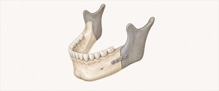
SSROSagittal Split Ramus Osteotomy
- Method
- Rigid fixation — fast, stable bone healing
- Best for
- Large movements or cases with a retruded chin
Three surgeries for the facial skeleton
Correction and restoration after previous surgery
Extraction · Implants · Liposuction · Injections
Before-and-after records and FAQs
Phone · SNS · Online form · Naver booking
ORTHOGNATHIC SURGERY
When both jaws find their rightful place,
your face looks most like you.


01 — PHILOSOPHY
By correcting two lines — the upper and lower jaws —
we restore both your bite and facial harmony.
02 — THE JOURNEY
※ Your exact plan is provided individually after a precise 3D-CT diagnosis
SURGERY
About 3 hours
ANESTHESIA
General anesthesia
STAY
2-day hospital stay
STITCHES
Stitches out after 2 weeks
RECOVERY
About 14 days
03 — CONCERNS
The feelings you carried in front of the mirror — we will listen to them first.

When a misaligned bite makes daily life uncomfortable
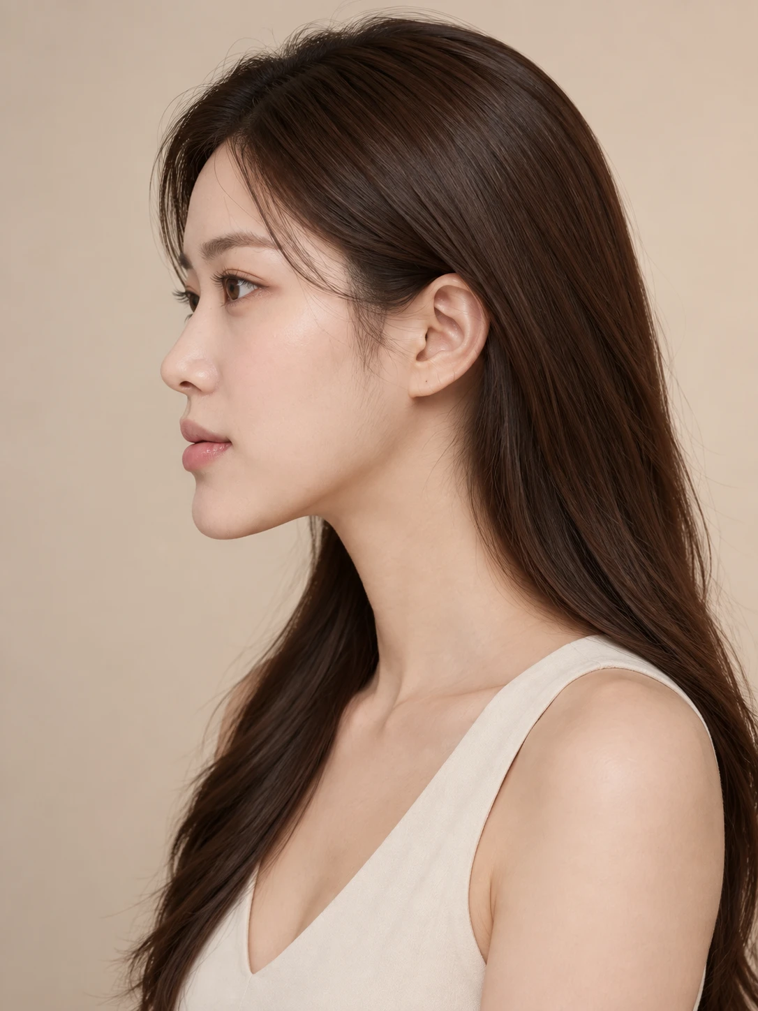
When your profile has always bothered you

When your mouth protrudes and your jawline looks faint

When the left and right sides look out of balance

When your face feels unusually long
04 — CASE STUDY
Select a concern to see tailored information.
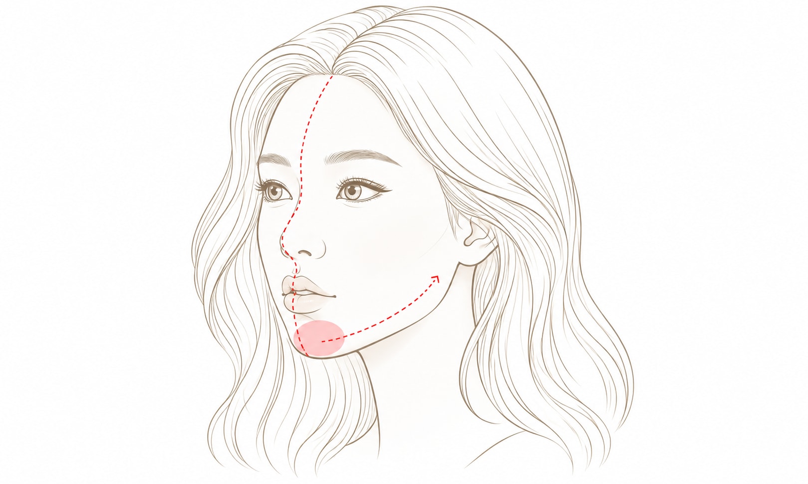
WHAT
The lower jaw protrudes forward, making the profile look strong
and chewing and speaking uncomfortable.
HOW
The lower jaw moves back and, if needed, the upper jaw moves forward.
Bite and profile are refined together.
Doctor’s comment
A mild underbite can give a mature impression.
Confirm whether surgery is truly needed with a precise 3D-CT diagnosis.
Osteotomy position and movement vary for each patient based on 3D-CT diagnosis
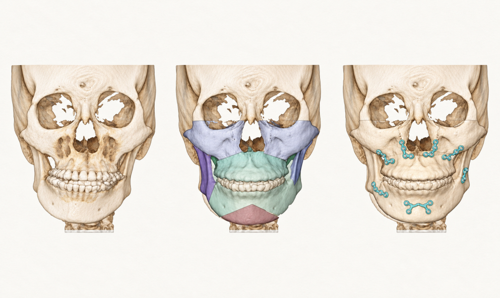
STEP 1
Osteotomy
Upper and lower jaw osteotomy following the 3D-CT plan
STEP 2
Movement · Fixation
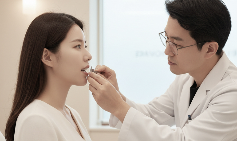
Moved in the direction suited to your type, then fixed
STEP 3
Bite · Line Check
Bite and profile checked through to the end
WHAT
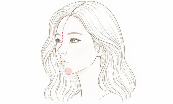
The lower jaw sits back or is small, so the jawline looks undefined and the mouth appears relatively protruded.
HOW
The lower jaw is advanced, combined with chin surgery (genioplasty) if needed — improving the chin and bite together. A prior evaluation of the jaw joint (TMJ) is essential.
Doctor’s comment
A mild retruded chin can give a youthful, soft impression. However, the approach differs depending on whether the issue is the chin alone or the jaw structure,
so we recommend confirming your condition through a precise diagnosis.
Osteotomy position and movement vary for each patient based on 3D-CT diagnosis
STEP 1
Osteotomy
Chin osteotomy following the 3D-CT plan
STEP 2
Advancement · Fixation
Lower jaw advanced, then fixed
STEP 3
Chin · Line Check
Chin position and profile checked
WHAT
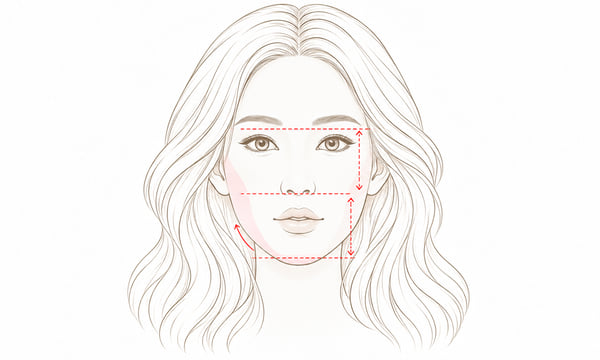
Vertical overgrowth of the upper jaw makes the mid and lower face look long, with excessive gum exposure.
HOW
Maxillary impaction reduces vertical length, and rotation of the lower jaw balances the proportions.
Doctor’s comment
A long face is not simply a face that looks long — it is often caused by vertical overgrowth of the upper jaw.
As gum exposure or bite problems often accompany it, a precise 3D-CT diagnosis is the most accurate way to confirm whether surgery is needed.
Osteotomy position and movement vary for each patient based on 3D-CT diagnosis
STEP 1
Maxillary Osteotomy (Impaction)
Reduces vertical length
STEP 2
Movement · Fixation
Fixation including lower jaw rotation
STEP 3
Proportion · Line Check
Vertical proportions and lines checked
WHAT
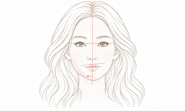
The upper and lower jaws are twisted or tilted to one side so the facial midline does not align — a condition in which one side has grown more than the other.
HOW
Rotation and tilt are corrected based on 3D-CT to align the midline and the occlusal plane.
Doctor’s comment
Facial asymmetry is not simply the two sides looking different — it is often caused by differences in jaw rotation and tilt.
The extent of surgery depends on the degree of asymmetry and any bite problems, so identifying the exact cause with a precise 3D-CT diagnosis is important.
Osteotomy position and movement vary for each patient based on 3D-CT diagnosis
STEP 1
3D-CT Analysis
Precise analysis of left-right deviation
STEP 2
Osteotomy · Rotational Correction
Fixed after axis rotation
STEP 3
Midline Check
Midline and occlusal plane checked
05 — METHOD
There is no single right answer. We choose based on your bone structure, nerve path and jaw joint condition.
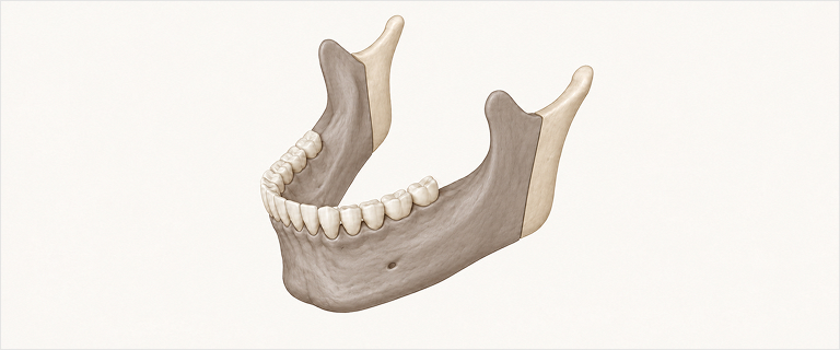
※ The final choice is made after analyzing jawbone thickness, nerve path and jaw joint condition with 3D-CT
06 — 3D DIGITAL SURGERY
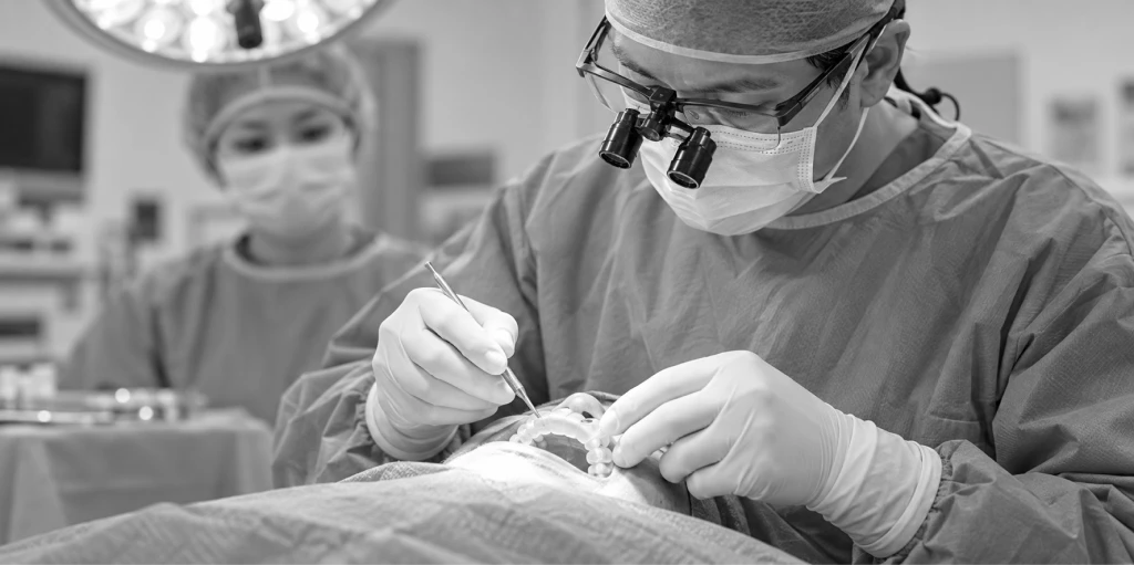
Surgical guides and custom plates are 3D-printed in advance, so surgery follows the plan exactly.
Reading bone structure and nerve paths
down to the millimeter
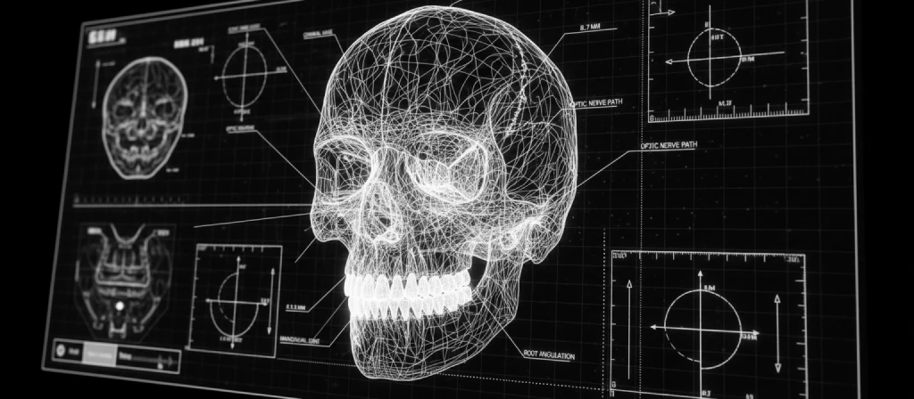
We perform the surgery
in a virtual space first
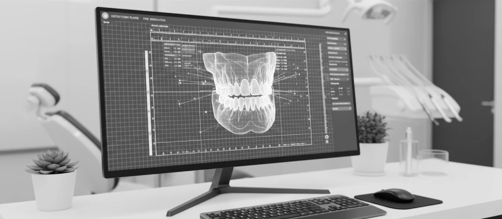
A guide that fits your bone structure,
made with 3D printing
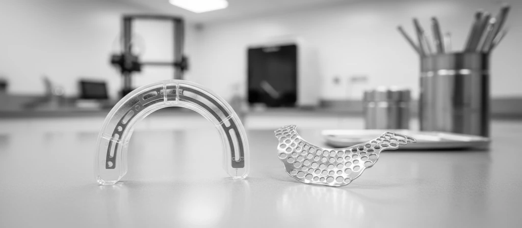
Exactly as planned,
without unnecessary movement
07 — DOCTOR
“ Turning hidden gems into jewels ”
Focusing wholly on one patient at a time,we seek the safest ideal outcome.
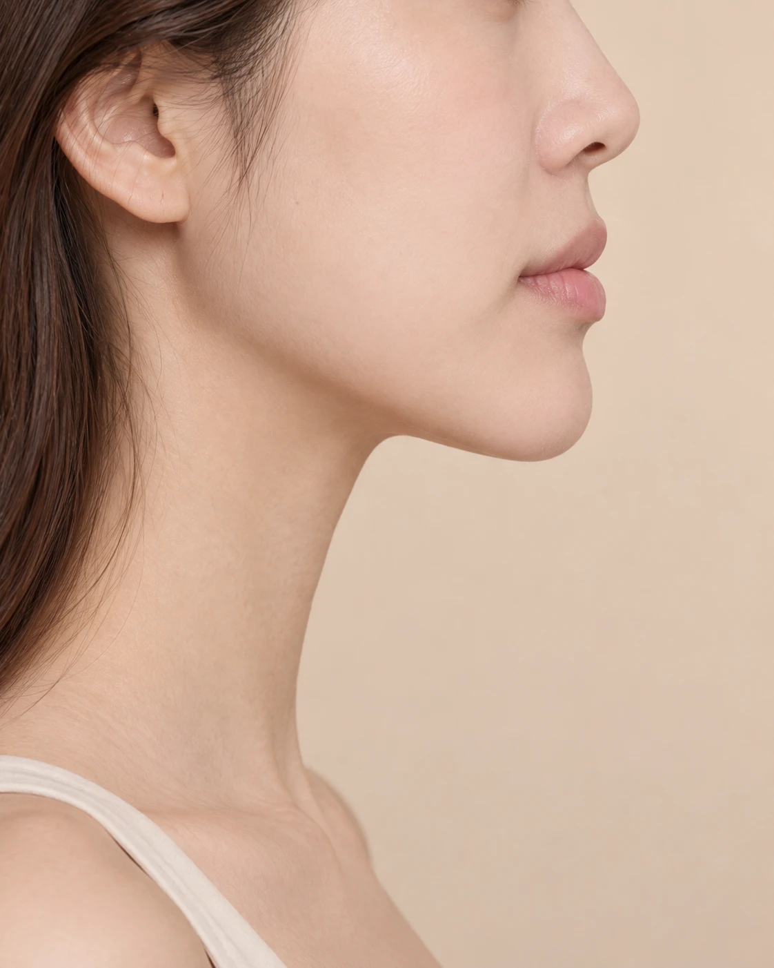
08 — BEFORE & AFTER
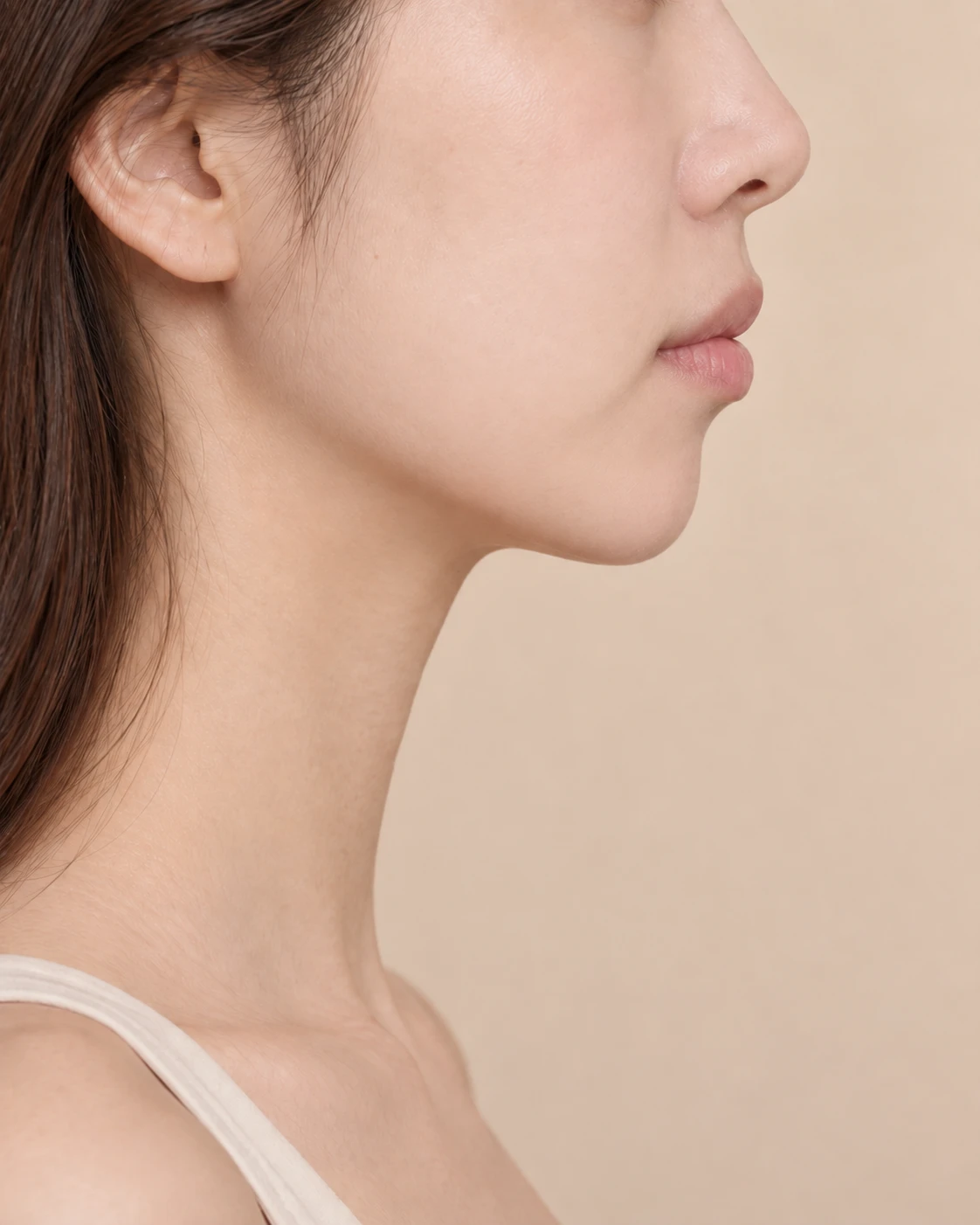
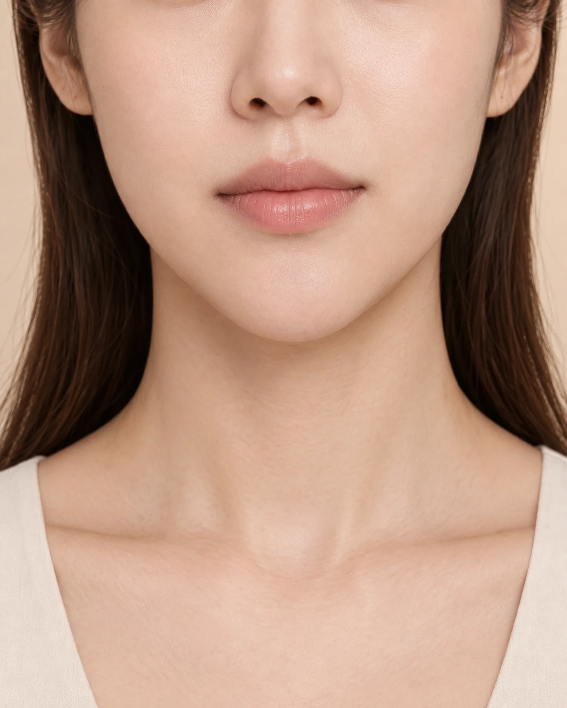
09 — QUESTIONS
For further questions,we will explain in detail during your consultation.
We decide after a precise skeletal analysis.
If it is possible, the overall treatment period can be noticeably shorter.
They can be planned together depending on your bone structure. Whether to combine them is decided by 3D-CT diagnosis.
We recommend removing fixation pins after the bone has healed. Details are provided on the pin removal page.
A twisted bite or axis is addressed with orthognathic surgery, while reshaping the bone itself is addressed with contouring surgery — the approaches differ.
When the lower jaw sits back, the mouth can look relatively protruded (retruded chin), so it must be distinguished from true protrusion. Since the two are treated differently, we recommend confirming your condition through profile analysis and a specialist’s diagnosis.
Cosmetic purposes are not covered, but coverage may apply when the surgery aims to improve function such as chewing or speech and meets the criteria. Eligibility is explained after examination and diagnosis.
